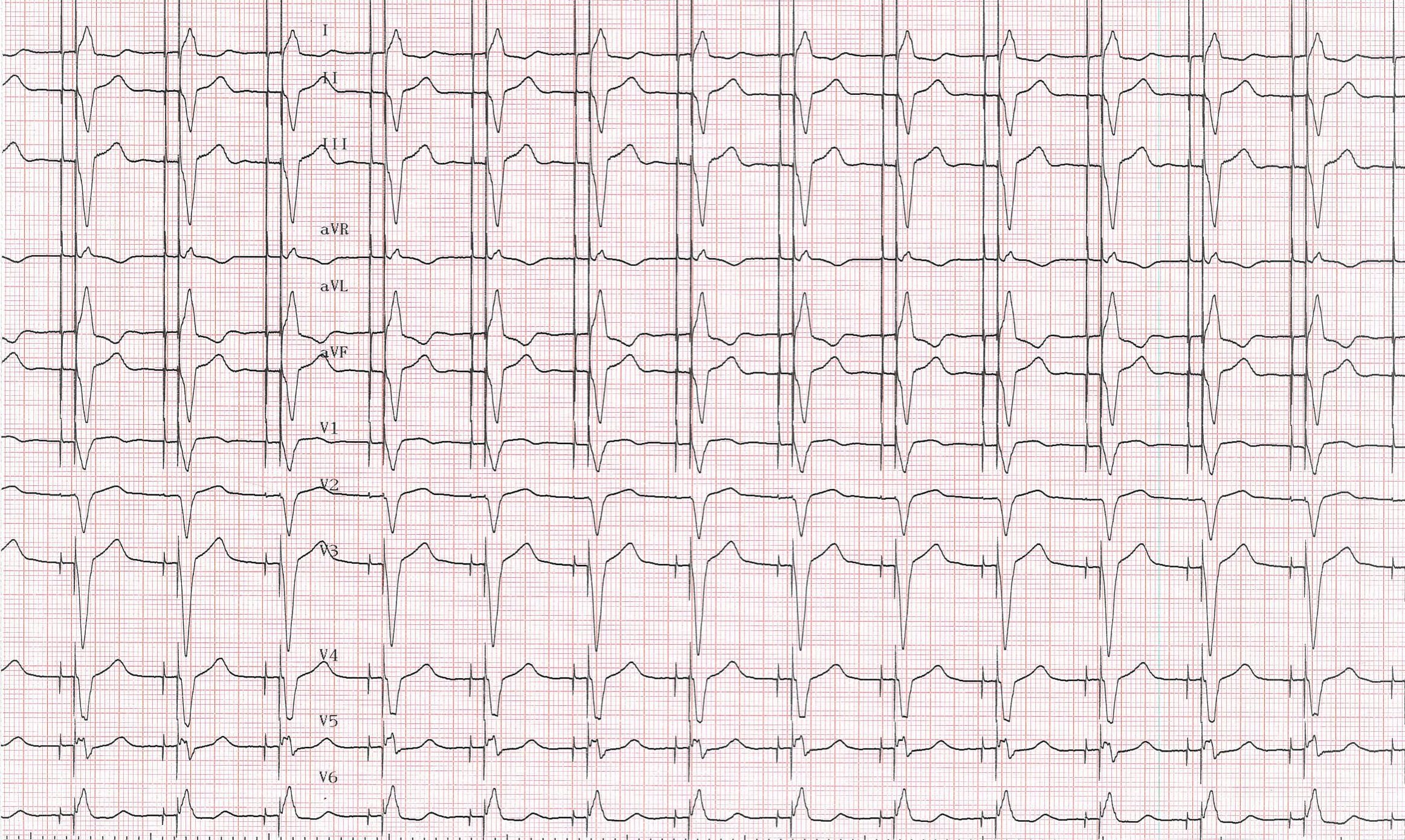
Patient
- 78-year-old man
- syncope and complete atrio-ventricular block
- Advisa DR MRI (Medtronic) dual-chamber pacemaker
- right atrial lead at the appendage, right ventricular lead at the apex
- 2 days after implantation, pacemaker control
ECG and dual chamber pacemaker
Time limit: 0
Case Summary
0 of 1 Questions completed
Questions:
Information
You have already completed the case before. Hence you can not start it again.
Case is loading…
You must sign in or sign up to start the case.
You must first complete the following:
Results
Case complete. Results are being recorded.
Results
0 of 1 Questions answered correctly
Time has elapsed
Categories
- Not categorized 0%
-
12 lead ECG
- sequential atrial and ventricular pacing
- apical right ventricular pacing with wide QRS complexes
- QRS complexes positive in DI, negative in inferior leads (DII, DIII, aVF)
- QRS complexes negative in V1 and positive in V6
Comments
- the surface ECG provides important information on lead position, ability to sense, to pace and to capture and is therefore determinant in pacemaker follow-up
- the challenges associated with interpreting the paced 12-lead electrocardiogram have greatly increased with the complexity of the most recent devices
- in patients implanted with a dual-chamber pacemaker, the ECG may exhibit features of atrial pacing, ventricular pacing or both; to correctly interpret a paced 12-lead ECG, a clinician needs to be aware of different rate and timing settings such as the pacing mode, the minimal rate, the maximal tracking rate, a variety of refractory periods, the AV delay …
- pacing artifacts may be hardly identifiable and careful analysis of the 12 leads may be required not to miss a spike preceding an atrial activity or a QRS complex; bipolar pacing configuration results in smaller pacing spike than unipolar pacing configuration
- the typical morphology of a right ventricular apical paced complex is that of left bundle branch block with superior axis (negative QRS complexes in the inferior leads); intermediate or inferiorly directed QRS complex axis are suggestive of leads positioned on the septum or in the outflow tract (dominant R wave in the inferior leads and qR, QR, or Qr complexes in leads I and aVL)
- ST segments and T waves are usually discordant with the QRS complex; inverted broad T waves may resemble ischemic T waves
- a right bundle branch block morphology suggests left ventricular pacing, whether intentional or not (lead placed inadvertently through a foramen ovale)
- the morphology of the paced atrial activity depends on lead placement; if the site of stimulation is near the sinus node, the morphology of the P waves resembles that of sinus beats; if the site of stimulation is closed to the coronary sinus, in the lower portion of the right atrium, the morphology of the atrial activity is inverted and negative in inferior leads
- 1
- Current
- Review / Skip
- Answered
- Correct
- Incorrect
-
Question 1 of 1
1. Question
On this ECG we can see the pacemaker in action, there is:
CorrectIncorrect