Patient
- 73-year-old woman
- syncope and complete atrio-ventricular block
- dual-chamber pacemaker
- chest X-ray 1 day after implantation
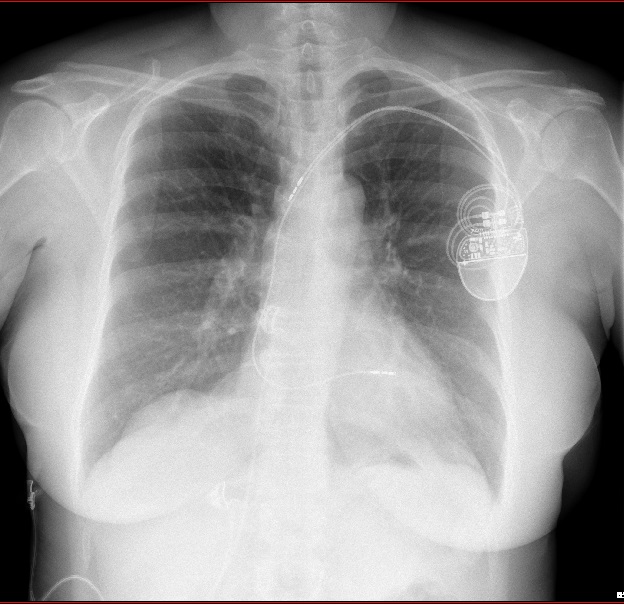
Chest X-ray: antero-posterior view
- right atrial lead dislodged into the superior vena cava
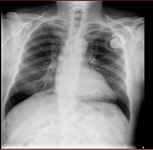
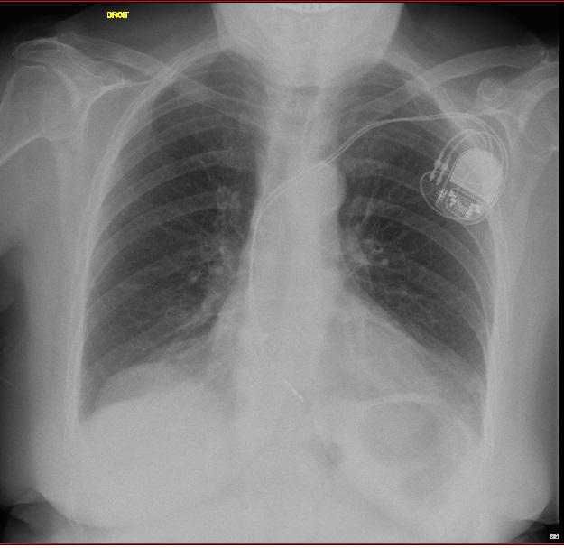
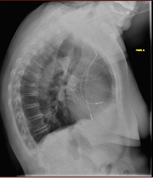
Now let’s look at some other X-rays.
lead dislodgement
Time limit: 0
Case Summary
0 of 2 Questions completed
Questions:
Information
You have already completed the case before. Hence you can not start it again.
Case is loading…
You must sign in or sign up to start the case.
You must first complete the following:
Results
Case complete. Results are being recorded.
Results
0 of 2 Questions answered correctly
Time has elapsed
Categories
- Not categorized 0%
-
Comments
- pacemaker leads are either equipped with tines (passive fixation) or with a screw (active fixation); both types of electrodes provide similar results in a long-term follow-up and the use of one or the other electrode type is mainly based on the implanter´s preference
- active fixation is accomplished by means of a screw tip, which can be visible on chest X-ray
- passive fixation leads are caught in the trabeculae lining the atrium or ventricle
- chest radiography is the preferred imaging modality to evaluate leads location, lead wire integrity and help in identifying several complications
- lead dislodgements have been classified as “macrodislodgement” versus “micro-dislodgement”, early versus late dislodgement
- macrodislodgement is radiographically evident whereas microdislodgement is not (harder to detect)
- early dislodgements occur within the first six weeks after implantation, and late dislodgements, after this period of time
- early dislodgements are much more frequent than late dislodgements and affect more frequently atrial leads
- 1
- 2
- Current
- Review / Skip
- Answered
- Correct
- Incorrect
-
Question 1 of 2
1. Question
Which lead is dislodged?
CorrectIncorrect -
Question 2 of 2
2. Question
There is dislodgement of the
CorrectIncorrect