Patient
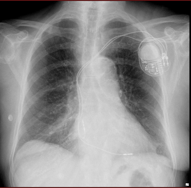
- 76-year-old woman
- syncope and complete atrio-ventricular block
- dual-chamber pacemaker
- chest X-ray 1 day after implantation
Chest X-ray: antero-posterior view
- right atrial lead placed via the left subclavian vein
- the right atrial lead is directed inferiorly and takes a J loop curve with the tip at the right atrial appendage
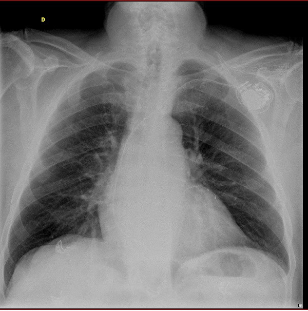
Now let’s see two other patients.
right atrial pacing site
Time limit: 0
Case Summary
0 of 2 Questions completed
Questions:
Information
You have already completed the case before. Hence you can not start it again.
Case is loading…
You must sign in or sign up to start the case.
You must first complete the following:
Results
Case complete. Results are being recorded.
Results
0 of 2 Questions answered correctly
Time has elapsed
Categories
- Not categorized 0%
-
Comments
- the conventional site for atrial pacing is the right atrial appendage because it is an easily accessible and stable location
- when the atrial lead is positioned at the appendage, the chest X-ray shows an inferiorly directed lead into the right atrium, with an anterior curve (“J loop”)
- pacing from the right atrial appendage in patients with pathological atrial myocardium prolongs the intra-atrial/inter-atrial conduction time and may increase the risk of atrial fibrillation
- alternative pacing sites within the right atrium (Bachmann’s bundle, interatrial septum, ostium of the coronary sinus or multiple atrial sites) have been investigated to prevent the occurrence of atrial fibrillation
- despite initial promising results with significant reduction of the total atrial activation time, the value of septal pacing or biatrial pacing in preventing atrial fibrillation has not been clearly demonstrated
- the lateral atrial wall is usually avoided because of the increased risk of perforation and of phrenic nerve capture
- 1
- 2
- Current
- Review / Skip
- Answered
- Correct
- Incorrect


